


Authors: Akiyuki Hiraga, Kazuho Kojima, Satoshi Kuwabara
Editor's Choice
Journal of the Neurological Sciences. REVIEW ARTICLE| VOLUME 461, 123045, JUNE 15, 2024
DOI: https://doi.org/10.1016/j.jns.2024.123045
Highlights
-
We analyzed clinical features of WE confirmed by low blood VB1 levels.
-
Corpus callosum and fornix were frequently involved in WE.
- MRI abnormalities were correlated with low blood vitamin B1 levels in WE.
- Note: Wernicke's encephalopathy, WE; Vitamin B1, VB1; magnetic resonance imaging, MRI.
Abstract
Purpose
Clinical features of Wernicke's encephalopathy (WE) confirmed strictly through the low blood vitamin B1 (VB1) levels are limited. This study aimed to analyse magnetic resonance imaging (MRI) findings, and clinical characteristics, in patients with WE who have confirmed low blood VB1 levels.
Methods
Clinical and laboratory records of 12 consecutive patients with WE admitted to our hospital during the past 11 years were reviewed. The WE diagnosis was confirmed based on low blood VB1 levels and the presence of at least one of the classical triad.
Results
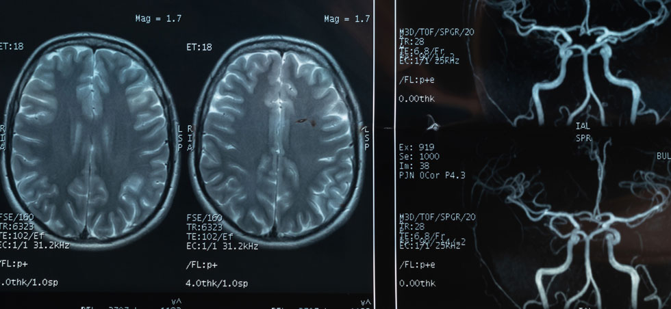
Ophthalmoplegia and nystagmus were recorded in 75% and 50% of the patients, respectively. Eleven of 12 patients presented with consciousness disturbance/memory loss. All patients experienced gait disturbances. Eight of the 12 patients exhibited MRI abnormalities at typical sites (the dorsal midbrain [n = 7], medial thalamus [n = 6], mammillary bodies [n = 5], and dorsal pons [n = 5]). Of the 12 patients, six showed abnormalities at atypical sites (the splenium of the corpus callosum [n = 4], fornix [n = 3], cerebral cortex [n = 2], cerebellar vermis [n = 2], and dorsal medulla [n = 1]). Patients with positive MRI abnormalities had significantly lower blood VB1 levels than those without abnormalities (9.5 vs. 16.0 ng/mL).
Conclusions
In cases of confirmed WE with low blood VB1 levels, the corpus callosum, fornix, and cerebral cortex were more frequently involved than in previous studies. MRI abnormalities at both typical and atypical sites were correlated with low blood VB1 levels in WE, suggesting that lower blood VB1 levels are associated with more severe brain damage in patients with WE.


Copyright © 2026 World Federation of Neurology. All rights reserved.
Web design by Tribal Systems