


Diagnosing progressive multifocal leukoencephalopathy: Positive predictive value of CSF JC virus quantitative PCR and importance of recognizing suggestive neuroimaging findings
Authors: Ryan Zhou, Kamala Sangam, Ana Cabrera, Fatimah AlMutawa, Aida Sivro, Felicia Roy, Michael Silverman, Manas Sharma, Adrian Budhram
Editor's Choice
Journal of the Neurological Sciences. REVIEW ARTICLE| VOLUME 469, 123379, February 15, 2025
DOI: https://doi.org/10.1016/j.jns.2024.123379
Highlights
- We found that a positive CSF JCV qPCR result had excellent PPV for PML in our cohort.
- All positives below the lowest quantifiable standard of the assay were true-positives.
- Differences in PPV across studies may relate to the population tested or assay used.
- All our positives had one or more neuroimaging findings reportedly suggestive of PML.
- Recognition of these suggestive neuroimaging findings helps facilitate PML diagnosis.
Abstract
Objective
To determine the positive predictive value (PPV) of CSF John Cunningham virus (JCV) quantitative PCR (qPCR) for progressive multifocal leukoencephalopathy (PML), and highlight neuroimaging findings reported to be suggestive of this disease.
Methods
We reviewed patients at London Health Sciences Centre with a positive CSF JCV qPCR result. Patients were classified as true-positive if they had a clinico-radiographic presentation compatible with PML and no more likely alternative diagnosis. The presence of suggestive neuroimaging findings was documented as supportive evidence of PML. The PPV of CSF JCV qPCR was calculated as the proportion of positive results that were classified as true-positives.
Results
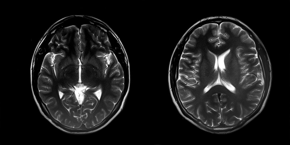
Eleven of 154 patients who underwent CSF JCV qPCR testing had a positive result (7 %). Median age was 60 years (range: 33–79 years) and 7/11 (64 %) were male. Nine of 11 (82 %) were overtly immunocompromised. Five of 11 (45 %) had a viral load below the lowest quantifiable standard (<4290 copies/ml). All had a clinico-radiographic presentation compatible with PML and no more likely alternative diagnosis, resulting in a PPV of 100 %. All had one or more suggestive neuroimaging findings that supported PML diagnosis (Milky Way sign/punctate pattern, 9; rim-and-core pattern, 7; T2/FLAIR mismatch, 6; shrimp sign, 4; SWI-hypointense rim, 2; across-the-pons sign, 1; barbell sign, 1).
Conclusions
We found that CSF JCV qPCR had high PPV for PML. All positives below the lowest quantifiable standard were true-positives. Our study affirms the diagnostic utility of this testing in clinical practice. Recognition of suggestive neuroimaging findings helps facilitate PML diagnosis.


Web design by Tribal Systems