


A look back at the relationship between electricity and the brain on the 100th anniversary of the first human EEG recording.
by Peter J. Koehler
This year marks the centennial of the first registration of a human electroencephalogram (EEG). Why is it that just a few hundred years ago physicians were still thinking in terms of animal spirits (spiritus animalis) flowing through cerebral ventricles and hollow nerves, when today we can create brain-computer interfaces to help disabled persons control their wheelchairs?
“Neurophysiology” Before the 18th Century
The doctrine of pneuma (spiritus in Latin, a very fine and volatile material principle of life) states that natural spirits (spiritus naturalis) arise in the liver, according to the physiology of Galen (129-c.199). These are converted into vital spirits (spiritus vitalis) after passing through tiny pores between the right and left parts of the heart. After passing through the rete mirabele — a vascular plexus of blood vessels at the base of the brain surrounding the pituitary gland that is present in mammals, but not in humans — they become animal spirits (spiritus animalis), flowing through the brain cavities and nerves. Greco-Roman medicine, with the even older humoral pathophysiology in addition to this pneuma doctrine, was influential for many centuries.
In the Middle Ages, the material part of the soul was still localized in the brain cavities — a system also known as ventricular or cell doctrine. It was assumed that in the first cell — our first and second ventricles — the information from the senses comes together.
In the 18th century, electric fish, specifically the electric eel, found in Dutch colonies in the West Indies — present-day Surinam and British Guiana — played a role in the realization that electricity is connected to nerve function. These fish can build up a potential of 700 to 800 volts, much more than the electric ray (torpedo), which had already been described in Ancient Greece.
Neurophysiology in the 19th Century
Finally, it was Emile Dubois Reymond (1818-1896) who was able to demonstrate the action potential (he called it “negative variation”) in a peripheral nerve with a sensitive galvanometer (1848). With this, he provided irrefutable proof that nerves were actually “electric,” which greatly influenced thinking about the etiology and treatment of nervous diseases. Shortly thereafter, the inventor of the ophthalmoscope (1851), Hermann von Helmholtz (1821-1894), was able to determine the velocity of the signal that propagates along the sciatic nerve of a frog.
Electrodiagnostics
Shortly thereafter, Richard Caton (1842-1926) in Liverpool was able to observe “feeble currents of varying direction” with electrodes on the cortex of a rabbit using a sensitive coil galvanometer combined with optical magnification. Several other researchers engaged in similar studies, including Adolf Beck (1863-1942) in Kraków. In 1890, Beck observed in laboratory animals that spontaneous fluctuations ceased after sensory stimulation. He also observed desynchronization of cortical activity upon sensory stimulation.
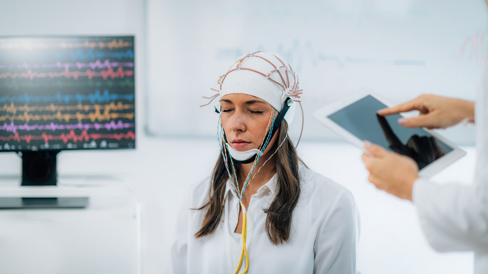
In 1901, Leiden physiologist Willem Einthoven (1860-1927) invented the string galvanometer, which shortly thereafter enabled him to record the ECG. He received the Nobel Prize for this in 1924. Hans Berger (1873-1941), who was interested in psycho-physical correlation through a telepathy (he did not use that term) incident, used this instrument — and later the Edelmann string galvanometer — for the registration of brain waves in humans. This year marks the centennial of his first registration from the human cortex in someone with a skull defect.
The discovery of EEG had a huge impact, both on science and the general public.
Brain Organ
Cybernetic theory-inspired physicist Edmond Dewan (b. 1931) — a friend of the pioneer in that field, the mathematician and philosopher Norbert Wiener (1894-1964) — designed a “brainwave control system” in the 1960s that allowed him to turn off a bedside lamp without using motor skills.
As an amateur organist, Dewan befriended experimental composer Alvin Lucier (1931-2021), who was inspired to create the first composition based on brainwaves. Music for Solo Performer was first performed in 1965. Sitting on a chair with his eyes closed, Lucier’s EEG was transmitted to numerous speakers scattered around the auditorium. Because the amplified alpha rhythm was below human audible range, the speakers were placed directly opposite various percussion instruments, which were then activated by vibration. As Lucier tried to refrain from mental activity, percussion sounds gradually emerged in the room. These then suddenly stopped again when he opened his eyes, engaged in mental exercises, or when his attention was drawn to sounds from the audience.
Brain-computer music interfaces became very popular. In 1995, when I attended a meeting of the European Club for the History of Neurology in Oslo, Norway, the opening ceremony took place in the Town Hall. The Norwegian composer Arne Nordheim (1931-2010), a pioneer of electronic music in Norway, produced music from signals routed to a computer from the EEG recording of a mother and her epileptic child. Today, such an experiment would probably raise eyebrows and ethical questions, but at the time, it was reported in Norwegian newspapers.
The sonification of brain waves took a new turn toward the late 1960s, when cybernetic theories and scientific breakthroughs gave rise to the field of biofeedback, in which biological processes were measured and fed back to the same individual to gain control over those processes. The sonification of EEG provided an interaction between neurophysiology and experimental music. Although initially applied by neurophysiologists as an adjunct visual EEG analysis, experimental composers turned to brainwave sonification to explore the sonic boundaries between mind and machine.
Brain-Computer Interfaces
During the past decades, the EEG signal became important as a diagnostic tool for communication with patients suffering from disorders of consciousness. In the field of rehabilitation of paralyzed patients, intracortical brain-computer interfaces are being used to restore movement and communication by decoding movement signals from the brain. Brain-computer interfaces enabled functional restoration of movement and communication, including robotic arm control, reanimation of paralyzed limbs through electrical stimulation, cursor control, translating attempted handwriting movements into text, and decoding speech.
EEG became a promising tool for bridging the gap between mind and machine, enabling the integration of mental and computer processes into one comprehensive system. Berger probably would have been thrilled.
Editor's Choice
World Neurology | May-June 2024, Volume 39, No. 3


Web design by Tribal Systems